Introduction
Durian (Durio zibethinus) is grown commercially across Malaysia, Thailand, Indonesia, Vietnam and the Philippines, and disease pressure is one of the most consistent constraints on orchard productivity in all five markets. A small number of pathogens, principally Phytophthora palmivora, account for the majority of yield-limiting disease in durian, but growers commonly encounter several other fungal pathogens that produce broadly similar symptoms and are frequently confused with one another in the field.
This article sets out the main durian diseases growers are likely to encounter, how to tell them apart, and the management principles supported by current research. It is intended as a diagnostic starting point, not a substitute for laboratory confirmation or a registered product label.
Quick Summary
- Phytophthora palmivora is the single most economically damaging durian pathogen in Southeast Asia, causing root rot, stem/patch canker and fruit rot (Singh et al., 2025).
- Disease losses attributed to Phytophthora across the region have been estimated at 20 to 25 percent of production, equivalent to roughly US$2.3 billion (Drenth & Guest, 2004, as cited in Singh et al., 2025).
- Leaf spot and blight symptoms can be caused by several distinct fungal genera, Colletotrichum, Phomopsis and Fusarium, that look similar to the untrained eye but require different management responses (Xu et al., 2025; Tongsri et al., 2016).
- Consistent management leans on orchard drainage, canopy hygiene and appropriately timed fungicide rotation rather than any single treatment (Kongtragoul et al., 2021).
Why This Matters
Phytophthora infection rates as high as 30 to 40 percent of trees have been documented in surveyed orchards in Penang and the southern Philippines (Singh et al., 2025). Because symptoms often begin below ground or inside the stem, trees can be significantly compromised before any visible canopy decline appears, which is why early, symptom-literate diagnosis matters commercially as well as agronomically.
Phytophthora Diseases: Root Rot, Stem Canker and Fruit Rot Phytophthora palmivora is an oomycete, not a true fungus, and is the dominant cause of root rot, stem or patch canker, and fruit rot in durian across Thailand, Malaysia, Indonesia, the Philippines and increasingly southern China (Dong et al., 2025; Singh et al., 2025). A second oomycete, Phytopythium vexans, and in some regions Pythium cucurbitacearum, produce overlapping symptoms and are frequently isolated alongside P. palmivora from the same orchard (Singh et al., 2025).
| Disease Phase | Key Symptoms | Typical Conditions |
| Root rot |
Fine feeder root loss, canopy thinning, dieback of terminal shoots, tree decline without an obvious trunk lesion |
Poorly drained soil, waterlogging after heavy rain |
| Stem / patch canker |
Dark, water-soaked lesions on the trunk, often with gum (resin) exudate; bark that peels back to reveal reddish-brown discoloured tissue |
Wounded bark, high humidity, splash dispersal from soil |
| Fruit rot | Brown, irregular lesions on the husk that expand rapidly, often starting at the fruit base or where fruit contacts soil or another fruit | Wet weather approaching harvest, fruit-to-soil or fruit-to-fruit contact |
Leaf Spot and Blight Diseases
Not every leaf lesion on a durian tree is Phytophthora. Several fungal genera cause leaf spot and blight independently of the oomycete diseases above, and distinguishing them matters because their biology and fungicide sensitivity differ.
| Pathogen | Typical Symptom Pattern | Notes |
| Colletotrichum spp. (anthracnose) | Circular to irregular brown lesions, sometimes with concentric rings; can extend to fruit anthracnose | Seven Colletotrichum species have been confirmed on durian leaves in newly surveyed plantations, most commonly C. fructicola and C. siamense (Xu et al., 2025) |
| Phomopsis durionis | Leaf spot with a distinct margin; the pathogen can also persist as a latent, symptomless infection before lesions appear |
First formally characterised on durian leaf tissue by Tongsri et al. (2016) |
| Fusarium spp. | Necrotic lesions on wounded and, for some species, unwounded leaves | Recently reported for the first time as a cause of durian foliar disease in newly expanding growing regions |
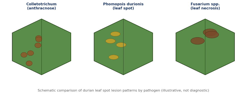
Figure 1. Schematic comparison of durian leaf spot lesion patterns across three fungal pathogen groups. ALT text: Illustrated comparison of durian leaf spot lesion patterns caused by Colletotrichum, Phomopsis and Fusarium Source: Original diagram created in-house from the cited literature (Xu et al., 2025; Tongsri et al., 2016); schematic and illustrative, not a substitute for photographic reference or laboratory diagnosis.
Diagnosis: Symptom to Confirmation
Field symptoms narrow the likely cause but rarely confirm it. The practical diagnostic sequence a grower or technician should follow is: observe the symptom pattern and its location on the tree, check the recent weather and drainage history of the block, rule out obvious non-disease causes such as mechanical damage or waterlogging stress, and, where the finding will change the management decision or the disease is spreading, send a sample for laboratory identification.
Laboratory confirmation matters most for canker and root rot symptoms, since Phytophthora, Phytopythium and Pythium all produce broadly similar lesions in the field but are identified definitively only through molecular or culture-based methods (Singh et al., 2025).
Differential Diagnosis: Ruling Out Look-Alikes
- Waterlogging stress alone can cause canopy thinning without any canker lesion; check for a lesion and gum exudate before assuming Phytophthora.
- Sunscald or mechanical bark injury can resemble early canker but lacks the internal reddish-brown discoloration typical of Phytophthora infection.
- Nutrient-related leaf yellowing is diffuse and follows a vein pattern, unlike the discrete lesions typical of Colletotrichum, Phomopsis or Fusarium leaf spot (see the Durian Doctor Nutrient Disorders article for deficiency-specific patterns).
Management
No single treatment reliably controls Phytophthora in durian; the research base supports an integrated approach combining cultural, chemical and, increasingly, biological measures (Singh et al., 2025).
- Orchard drainage: improve surface and subsurface drainage in root rot–prone blocks, since waterlogging is consistently associated with higher infection rates.
- Canopy and trunk hygiene: remove and destroy infected fruit and cankered tissue promptly rather than leaving inoculum in the orchard.
- Fungicide rotation: where chemical control can be recommended for use in durian farming in your market, growers should follow the specific product label for rate, timing and pre-harvest interval, and rotate modes of action; monitoring in Thailand has documented reduced sensitivity of some P. palmivora populations to metalaxyl, underscoring the importance of rotation rather than repeated single-mode-of-action use (Kongtragoul et al., 2021).
- Biological and alternative approaches: organic soil amendments and composted mulches have been shown to support beneficial soil microbial activity that suppresses Phytophthora survival, and are best used as a complement to, not a replacement for, cultural and chemical measures (Singh et al., 2025).
- Postharvest handling: minimising fruit contact with soil during harvest, and cooling fruit promptly after picking, reduces postharvest fruit rot incidence and severity.
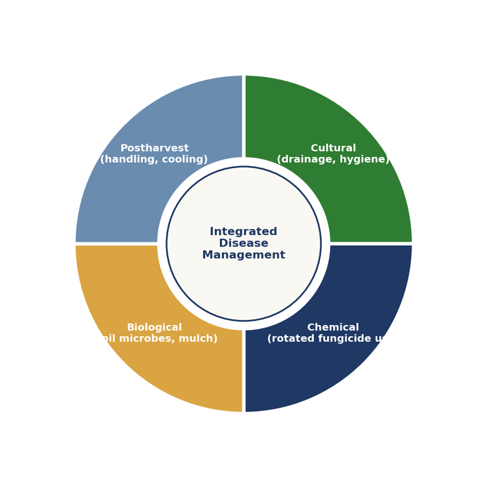
Figure 2. Integrated management cycle for durian Phytophthora diseases. ALT text: Diagram showing cultural, chemical, biological and postharvest control working together for durian Phytophthora management Source: Original diagram created in-house based on the cited literature.
Prevention
- Select planting sites with good natural drainage and avoid low-lying, waterlogging-prone ground where possible.
- Avoid mechanical wounding of trunks and exposed roots during orchard operations, since wounds are common infection points.
- Maintain a regular orchard scouting schedule through the wet season, when Phytophthora spread is most active.
- Where a block has a known disease history, prioritise it for closer monitoring and earlier intervention in subsequent seasons.
Practical Farm Checklist
| Check | Frequency |
| Inspect trunk base for gum exudate or discoloured bark | Monthly, weekly during wet season |
| Inspect canopy for dieback or thinning not explained by other stress | Monthly |
| Review block drainage after heavy rainfall events | After each major rain event |
| Remove and destroy fallen or infected fruit promptly |
Ongoing during fruiting season |
| Rotate fungicide mode of action between applications | Per spray programme |
Key Takeaways
- Phytophthora palmivora is the priority disease to manage in most durian-growing regions, but is not the only cause of canker, rot or leaf spot symptoms.
- Symptom location and pattern narrow the diagnosis; laboratory confirmation is warranted for spreading or high-value cases.
- Integrated management, drainage, hygiene, rotated fungicide use and biological support, consistently outperforms any single-tactic approach in the published literature.
- Product recommendations must always be checked against the current label and local registration status for your market.
Frequently Asked Questions
What is the most serious durian disease?
Phytophthora palmivora is generally regarded as the most economically significant durian pathogen across Southeast Asia, responsible for root rot, stem canker and fruit rot, with regional losses estimated at 20 to 25 percent of production (Drenth & Guest, 2004, as cited in Singh et al., 2025).
How can I tell Phytophthora canker apart from mechanical bark damage?
Phytophthora canker typically shows a water-soaked, dark lesion with gum exudate and reddish-brown discoloration beneath the bark when scraped back. Mechanical injury lacks this internal discoloration and usually has a clean, defined wound margin.
Is leaf spot always caused by the same pathogen?
No. Durian leaf spot can be caused by Colletotrichum, Phomopsis durionis, Fusarium species and others, which look similar in the field but differ in biology and treatment response. Where the diagnosis will change management, laboratory identification is recommended.
Can fungicides alone control Phytophthora in durian?
Fungicides are one part of an integrated programme. Reduced sensitivity to some commonly used actives has been documented in surveyed Thai populations, which is why rotation, drainage and orchard hygiene remain essential alongside chemical control (Kongtragoul et al., 2021).
When should I send a sample for laboratory testing?
Laboratory confirmation is most useful when a canker or root rot symptom is spreading, when previous treatment has not resolved the problem, or when the finding will materially change the management or replanting decision.
References
Dong, X., Wang, Y., Liu, Z., Feng, X., Guo, L., Deng, H., & colleagues. (2025). Phytophthora palmivora as a causal pathogen of stem canker and fruit rot of Durio zibethinus in China. Journal of Phytopathology. https://doi.org/10.1111/jph.70089
Drenth, A., & Guest, D. I. (2004). Diversity and management of Phytophthora in Southeast Asia. Australian Centre for International Agricultural Research (ACIAR) Monograph 114.
Kongtragoul, P., Ishikawa, K., & Ishii, H. (2021). Metalaxyl resistance of Phytophthora palmivora causing durian diseases in Thailand. Horticulturae, 7(10), 375. https://doi.org/10.3390/horticulturae7100375
Singh, A., Chow, C., Nathaniel, K., Yap, L. V., Javad, S., & Jabeen, K. (2025). Management of Phytophthora and Phytopythium oomycete diseases in durian (Durio zibethinus). Crop Protection, 190, 107086. https://doi.org/10.1016/j.cropro.2024.107086
Tongsri, V., Songkumarn, P., & Sangchote, S. (2016). Leaf spot characteristics of Phomopsis durionis on durian (Durio zibethinus Murray) and latent infection of the pathogen. Acta Universitatis Agriculturae et Silviculturae Mendelianae Brunensis, 64(1), 185–193.
Xu, H., Zhang, Z., Zhou, Z., West, J. S., Wang, J., & Cao, X. (2025). Colletotrichum species associated with durian (Durio zibethinus Murray) in Hainan, China. Crop Protection, 188, 107034. https://doi.org/10.1016/j.cropro.2024.107034